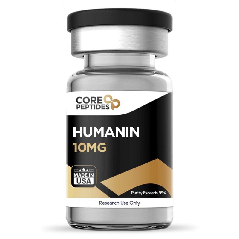
Humanin – 10MG
$147.00
Humanin is a short mitochondrial-derived peptide encoded in the 16S rRNA gene. Content size is 10mg, in the form of lyophilized powder. Peptides are sold for laboratory research use only.
Purity: 99%+ by HPLC (third-party tested)
Tested by: Vanguard Laboratory, see Certificate of Analysis
SKU: P-HUMANIN-10
Discount per Quantity
| Quantity | Discount | Price |
|---|
| 5 - 8 | 5% | $139.65 | |
| 9 + | 10% | $132.30 |
What Is Humanin?
Humanin is a short natural peptide with suggested potential in cell metabolism and inflammation response.(3) Researchers have suggested that Humanin is encoded by a gene within the mitochondrial genome (the 16S ribosomal RNA gene).(4) Researchers further posit that the peptide's length may depend on the location of its synthesis. The peptide may contain 21 amino acids if synthesized inside the mitochondria, whereas it may contain 24 amino acids if synthesized outside the mitochondria but inside the cytosol.(5) Both of these peptides exhibit potential biological activity.
Mitochondria are considered the powerhouses of the cell, and are formed from the engulfment of individual prokaryotes. Eventually, the eukaryotes appear to engulf the prokaryotic (single-celled) organism, and the prokaryotes form an endosymbiotic relationship with the host cell and gradually develop into mitochondria.(1)
Mitochondria are considered to be responsible for several vital cellular activities, including energy production, regulation of apoptosis, hemostasis, and formation of heme proteins, among several other functions. All these functions appear to be regulated by mitochondria via their communications to the cell through several retrograde signals. These signals may be encoded in the nuclear genome, possibly due to its prokaryotic origin.(2)
A small peptide called Humanin is derived from a gene present in the mitochondrial genome. Since this genome appears to play an important role, this peptide has been explored for its potential action in several biological functions.
How Is Humanin Thought to Work?
The peptide appears to exert potential actions via binding with intracellular molecules and cell membrane receptors, possibly inducing cytoprotective and/or neuroprotective functions.(9) Researchers suggest that Humanin may bind with the Bcl-2-associated X protein (also called Bax protein). Bax protein is considered to play a vital role in cellular death (apoptosis). Upon binding with the inactive form of the Bax protein, Humanin may inhibit the changes in the Bax protein and potentially thereby prevent cellular apoptosis.(9)
Apart from research into its possible interaction with Bax, Humanin studies suggest the peptide may also bind with other intracellular molecules, such as actinin-4 and phosphoprotein 8, which are both involved in cellular apoptosis. Upon binding with these proteins, Humanin may induce cytoprotective actions. Researchers suggest Humanin may bind with two G protein-coupled peptide receptors, namely FPRL-1 and FPRL-2 receptors, which are considered to be involved with neurological function.(9) By binding with this receptor, Humanin may potentially prevent amyloid β binding with the FPRL-1 and FPRL-2 receptors, which may mitigate some instances of neurological degradation.
Humanin Specifications
| Name | Humanin |
| Other known names | HN, Humanin (human) |
| Sequence | Met-Ala-Pro-Arg-Gly-Phe-Ser-Cys-Leu-Leu-Leu-Leu-Thr-Ser-Glu-Ile-Asp-Leu-Pro-Val-Lys-Arg-Arg-Ala (24 amino acids) |
| Molecular formula | C119H204N34O32S2 |
| Molecular weight | 2687.3 g/mol |
| CAS number | 330936-69-1 |
| Synthesis | Solid-phase peptide synthesis, US |
| Form | Lyophilized powder |
| Amount per vial | 10 mg |
| Purity | >99% (HPLC) |
| Storage | −20 °C, dry, protected from light |
| SKU | P-HUMANIN-10 |
What Does Research Show About Humanin?
How May Humanin Affect Mitochondrial Function in Research?
Key finding: In retinal pigment epithelial cells under oxidative stress (tert-butyl hydroperoxide), Humanin inhibited reactive oxygen species formation, restored mitochondrial bioenergetics, increased mitochondrial DNA copy number and protected the cells from oxidative stress-induced death and senescence.(10)
Mitochondria appear to be susceptible to reactive oxygen species (ROS), and the presence of such ROS may reduce its functioning. Studies in Humanin mechanisms of action suggest that the peptide may inhibit these reactive oxygen species to some degree, thereby mitigating mitochondria degeneration.(10) One study was produced wherein the researchers attempted to explore the peptide's action on retinal pigment epithelial cells. These cells were isolated and introduced to tert-butyl hydroperoxide to exert oxidative stress in the cells. Some of these cells were then presented with Humanin peptide for 24 hours. When examined, the cells exposed to Humanin reportedly inhibited the formation of tert-butyl hydroperoxide-induced reactive oxygen species. The researchers suggested the peptide may have restored the bioenergetics in the retinal pigment epithelial cells and increased the functioning of mitochondria.
Is Humanin Linked to Cellular Longevity in Research Models?
Key finding: In lab models with altered GH/IGF-I signaling, Humanin levels correlated negatively with GH–IGF axis activity and positively with lifespan: long-lived, GH-deficient (Ames) models had elevated Humanin levels, while short-lived GH-transgenic models had reduced levels.(11)
Studies(11) were conducted on several murine models to determine the relation between Humanin with growth hormone (GH) and insulin-like growth factor 1 (IGF-1). In GH-transgenic murine models, the levels of growth hormone and IGF-1 appeared to be extremely high, which the researchers suggested led to increased body size, accelerated cell aging, and reduced life span. Researchers reported that the levels of Humanin naturally present in the murine models were extremely low, about 70% lower than the control.
Another set of murine models had undetectable levels of GH and IGF-1, along with a reported 40% increase in the Humanin levels. These murine models showed an increased life span compared to normal murine models. These results supported the researchers' hypothesis that Humanin may be negatively correlated to GH and IGF-1 levels and directly correlated to cellular longevity.
What Does Research Show About Humanin and Neurological Deterioration?
Key finding: In cultured cortical neurons, preincubation with Humanin preserved cell viability, reduced oxidative stress, maintained PP2A activity and blocked tau over-phosphorylation at Ser199/202, Ser396 and Thr231 after calyculin A exposure.(14)
A study(12) was conducted on 9-month-old murine models, all of which reportedly possessed high levels of amyloid proteins. These amyloid chemicals are indicative of some neurological deterioration. Some murine models were presented with a Humanin analog (HNG), while others were given a placebo. Following the study, the researchers suggested that the placebo models showed impaired memory and poor learning skills. In contrast, the murine models presented with a Humanin analog for 3 months reportedly exhibited significantly improved learning ability and enhanced memory.
In another study,(13) murine models with experimentally induced impairment in neurological functioning were exposed to Humanin and a similarly structured peptide, PAGA, to determine the potential of both peptides on the brain function of the murine models. Both the peptides showed some modest improvement in the impairment of the brain, as suggested by the researchers.
Another study explored the potential neuroprotective action of Humanin on neurotoxicities induced by Calyculin A in cortical neurons as a model of exposure to neurotoxins. The researchers focused on the potential capability of the peptide to safeguard cortical neurons from Calyculin A-induced neurotoxic action. In this investigation, cortical neurons were preincubated with Humanin to examine its protective role against neurotoxicity. The methodologies utilized in this research included employing 3-(4,5-Dimethylthiazol-2-yl)-2,5-diphenyltetrazolium bromide (MTT), lactate dehydrogenase (LDH), and Calcein-AM assays to evaluate the extent of neural damage. Furthermore, caspase 3 signaling and TUNEL assays were conducted to assess neural apoptosis. The study also used Western blot analysis to detect potential expressions of phosphorylated tau, a protein associated with neurodegenerative diseases. Additionally, the study measured the apparent contents of malondialdehyde (MDA) and superoxide dismutase (SOD), and the activity of protein phosphatase 2A (PP2A), which are potential indicators of oxidative stress in neurons.
The results from these investigations posited that preincubation with Humanin may preserve cell viability and potentially protect neurons from Calyculin A-induced damage. Notably, the peptide appeared to alleviate oxidative stress in the neurons and preserved the activity of PP2A, an enzyme involved in dephosphorylating tau proteins. Humanin also may have inhibited the over-phosphorylation of tau at specific sites (Ser199/202, Ser396, and Thr231), which are considered critical to the pathological changes observed in neurodegenerative diseases. From these findings, it was inferred that Humanin potentially offered protective action against neurotoxicities induced by harmful agents like Calyculin A.(14)
What Does Research Show About Humanin and Insulin Resistance?
Key finding: In clamp studies in lab models, continuous exposure of the brain to Humanin significantly improved overall insulin sensitivity through hypothalamic STAT-3 signaling; blocking hypothalamic STAT-3 cancelled the effect.(17)
To determine the potential of Humanin on glucose tolerance, a study(15) was conducted on nonobese diabetic murine models. When a group of these models were presented with Humanin, it appeared to restore to some degree the levels of glucose tolerance within 6 weeks. Furthermore, Humanin also potentially delayed the onset of diabetes in the murine models presented with the peptide for 20 weeks. Researchers suggested that following the study results, Humanin may exhibit some action in insulin resistance.
In another study,(16) 12-week-old murine models were subjected to a 60% high-fat diet and were presented with Humanin for 4 weeks. After the study, there appeared to be no difference in the food intake. However, the weight gain had reportedly been reduced by about 20%. Furthermore, there appeared to be a high expenditure of energy, decreased fasting glucose levels, and increased insulin levels.
Another study also explored the potential role of Humanin in influencing insulin sensitivity. The investigation suggested that continuous exposure of Humanin to the central nervous system of murine models appeared to enhance overall insulin sensitivity. This improvement in insulin action seemed to be linked to the activation of STAT-3 (Signal Transducer and Activator of Transcription 3) signaling within the hypothalamus. However, this action was negated when hypothalamic STAT-3 was co-inhibited, suggesting a potential role of this signaling pathway in mediating Humanin actions on insulin sensitivity. Furthermore, centrally-acting Humanin also appeared to improve insulin resistance in peripheral tissues such as liver cells. Ultimately, this appeared to result in reduced blood glucose levels in the murine models.(17)
Could Humanin Protect Cells Under Hypoxia in Research?
Key finding: In RGC-5 retinal ganglion cells, Humanin significantly reduced the loss of viability and the apoptosis caused by the hypoxia-mimetic compound cobalt chloride.(18)
A study(18) was conducted in which the isolated retinal cells were exposed to cobalt chloride, which appeared to induce hypoxia, leading to cell apoptosis. When the hypoxia-induced cells were presented with Humanin, researchers reported that the peptide appeared to reverse the impact of cobalt chloride and protect the cell from reduced oxygen levels.
Additional studies (19) have suggested that Humanin may possibly increase metabolic activity and, thereby, cell survival rates in the event of lymphocyte death, which has some implications in ischemia.
What Does Research Show About Humanin and Ischemia?
Key finding: In lab models of focal cerebral ischemia/reperfusion, the potent Humanin analog HNG, given into the brain ventricles 30 minutes before ischemia, reduced infarct volume from 56.2% to 26.1%.(20)
This study(20) was conducted to further examine the neuroprotective potential of Humanin in the presence of cerebral ischemia. In this study, murine models were experimentally induced with cerebral artery occlusion. Murine models were presented with low concentrations of Humanin for 30 minutes and then, following the procedure, were reintroduced to Humanin at 0, 2, 4, and 6 hours after ischemia. Other murine models were solely presented with Humanin one hour before ischemia. It was suggested that continual introduction to the peptide appeared to reduce the ischemia volume by almost 30%. Humanin presence following ischemia reportedly further reduced the ischemic impact.
Another study on Humanin investigating its potential in mitigating ischemia in cells suggests that the peptide may be capable of reducing infarct size in large animal models.(21) In this notable study, researchers suggested that Humanin may exhibit a potential to significantly reduce infarct size by 41% in a large animal model when introduced alongside standard reperfusion intervention after an ischemic period of 60 minutes, followed by 48 hours of reperfusion. However, it is important to note that the beneficial action of Humanin was not observed when the ischemic period was extended to 75 minutes, followed by the same duration of reperfusion, highlighting a potentially critical window for Humanin. The study also highlights that Humanin has been implicated in various protective roles across multiple organs, attributed to the potential to modulate oxidative stress, regulate insulin sensitivity, and inhibit apoptotic signaling, among other actions.
What Other Effects of Humanin Have Been Studied?
Key finding: In lab models, Humanin delivered into the brain ventricles showed anxiolytic-like activity in the elevated plus-maze test, an effect blocked by the FPR2 antagonist WRW4.(22)
Studies(22) have suggested that Humanin may bind with the FPR2 receptor in the brain, producing possible anxiolytic action. Consequently, the researchers of this study suggested that Humanin may potentially reduce anxiety symptoms.
In this study,(23) murine carcinogenesis models were exposed twice weekly with an antineoplastic agent (used in cancer cases) and Humanin to explore their potential impact on the carcinogenic cells. While the control agent appeared to induce cell apoptosis, the combination of Humanin and the agent exhibited some potential, somewhat reversing the apoptosis of the healthy cells.
Humanin Certificate of Analysis
| Batch / lot | C73627 |
| Test date | 4/2/2026 |
| Testing laboratory | Vanguard Laboratory |
| Method | HPLC-UV/VIS (purity) |
| Purity result | >99.80% |
| Full report | Download the Humanin COA (PDF) |
Humanin peptide is available for research and laboratory purposes only. We offer free shipping in the US for orders over $200. Please review our Terms and Conditions before ordering.
Humanin Frequently Asked Questions
What is Humanin?
Humanin is a short peptide encoded in the mitochondrial genome (the 16S ribosomal RNA gene). Researchers suggest it contains 24 amino acids when made in the cytosol and 21 when made inside the mitochondria.
How is Humanin thought to protect cells?
Researchers suggest Humanin may bind the Bax protein and prevent its activation, and may also act on receptors such as FPRL-1 and FPRL-2, which could help protect cells from apoptosis.
What is the molecular weight of Humanin?
Humanin has the molecular formula C119H204N34O32S2 and a molecular weight of approximately 2687.3 g/mol.
How should Humanin be stored?
Lyophilized Humanin should be stored at −20 °C, dry and protected from light.
Is Humanin third-party tested?
Yes. The current batch (C73627) was tested by a third-party laboratory and showed >99% purity by HPLC. Its certificate of analysis is available on this page.
References:
- Nature Education. (n.d.). The origin of mitochondria and chloroplasts. Scitable by Nature Education. https://www.nature.com/scitable/content/the-origin-of-mitochondria-and-chloroplasts-14747702/
- Lee, C., Yen, K., & Cohen, P. (2013). Humanin: a harbinger of mitochondrial-derived peptides? Trends in endocrinology and metabolism: TEM, 24(5), 222–228. https://doi.org/10.1016/j.tem.2013.01.005
- Gong, Z., Tas, E., & Muzumdar, R. (2014). Humanin and age-related diseases: a new link? Frontiers in endocrinology, 5, 210. https://doi.org/10.3389/fendo.2014.00210
- Hashimoto, Y., Niikura, T., Tajima, H., Yasukawa, T., Sudo, H., Ito, Y., Kita, Y., Kawasumi, M., Kouyama, K., Doyu, M., Sobue, G., Koide, T., Tsuji, S., Lang, J., Kurokawa, K., & Nishimoto, I. (2001). A rescue factor abolishing neuronal cell death by a wide spectrum of familial Alzheimer's disease genes and Abeta. Proceedings of the National Academy of Sciences of the United States of America, 98(11), 6336–6341. https://doi.org/10.1073/pnas.101133498
- Yen, K., Lee, C., Mehta, H., & Cohen, P. (2013). The emerging role of the mitochondrial-derived peptide humanin in stress resistance. Journal of molecular endocrinology, 50(1), R11–R19. https://doi.org/10.1530/JME-12-0203
- Gong, Z., Tas, E., & Muzumdar, R. (2014). Humanin and age-related diseases: a new link? Frontiers in endocrinology, 5, 210. https://doi.org/10.3389/fendo.2014.00210
- Guo, B., Zhai, D., Cabezas, E., Welsh, K., Nouraini, S., Satterthwait, A. C., & Reed, J. C. (2003). Humanin peptide suppresses apoptosis by interfering with Bax activation. Nature, 423(6938), 456–461. https://doi.org/10.1038/nature01627
- Ikonen, M., Liu, B., Hashimoto, Y., Ma, L., Lee, K. W., Niikura, T., Nishimoto, I., & Cohen, P. (2003). Interaction between the Alzheimer's survival peptide humanin and insulin-like growth factor-binding protein 3 regulates cell survival and apoptosis. Proceedings of the National Academy of Sciences of the United States of America, 100(22), 13042–13047. https://doi.org/10.1073/pnas.2135111100
- Gong, Z., Tas, E., & Muzumdar, R. (2014). Humanin and age-related diseases: a new link? Frontiers in endocrinology, 5, 210. https://doi.org/10.3389/fendo.2014.00210
- Sreekumar, P. G., Ishikawa, K., Spee, C., Mehta, H. H., Wan, J., Yen, K., Cohen, P., Kannan, R., & Hinton, D. R. (2016). The mitochondrial-derived peptide humanin protects RPE cells from oxidative stress, senescence, and mitochondrial dysfunction. Investigative ophthalmology & visual science, 57(3), 1238–1253. https://doi.org/10.1167/iovs.15-17053
- Lee, C., Wan, J., Miyazaki, B., Fang, Y., Guevara-Aguirre, J., Yen, K., Longo, V., Bartke, A., & Cohen, P. (2014). IGF-I regulates the age-dependent signaling peptide humanin. Aging cell, 13(5), 958–961. https://doi.org/10.1111/acel.12243
- Zhang, W., Zhang, W., Li, Z., Hao, J., Zhang, Z., Liu, L., Mao, N., Miao, J., & Zhang, L. (2012). S14G-humanin improves cognitive deficits and reduces amyloid pathology in the middle-aged APPswe/PS1dE9 mice. Pharmacology, biochemistry, and behavior, 100(3), 361–369. https://doi.org/10.1016/j.pbb.2011.09.012
- Krejcova, G., Patocka, J., & Slaninova, J. (2004). Effect of humanin analogues on experimentally induced impairment of spatial memory in rats. Journal of peptide science, 10(10), 636–639. https://doi.org/10.1002/psc.569
- Zhao, J., Zeng, Y., Wang, Y., Shi, J., Zhao, W., Wu, B., & Du, H. (2021). Humanin protects cortical neurons from calyculin A-induced neurotoxicities by increasing PP2A activity and SOD. The International journal of neuroscience, 131(6), 527–535. https://doi.org/10.1080/00207454.2020.1769617
- Hoang, P. T., Park, P., Cobb, L. J., Paharkova-Vatchkova, V., Hakimi, M., Cohen, P., & Lee, K. W. (2010). The neurosurvival factor Humanin inhibits beta-cell apoptosis via signal transducer and activator of transcription 3 activation and delays and ameliorates diabetes in nonobese diabetic mice. Metabolism: clinical and experimental, 59(3), 343–349. https://doi.org/10.1016/j.metabol.2009.08.001
- Gong, Z., Su, K., Cui, L., Tas, E., Zhang, T., Dong, H. H., Yakar, S., & Muzumdar, R. H. (2015). Central effects of humanin on hepatic triglyceride secretion. American journal of physiology. Endocrinology and metabolism, 309(3), E283–E292. https://doi.org/10.1152/ajpendo.00043.2015
- Muzumdar, R. H., Huffman, D. M., Atzmon, G., Buettner, C., Cobb, L. J., Fishman, S., Budagov, T., Cui, L., Einstein, F. H., Poduval, A., Hwang, D., Barzilai, N., & Cohen, P. (2009). Humanin: a novel central regulator of peripheral insulin action. PloS one, 4(7), e6334. https://doi.org/10.1371/journal.pone.0006334
- Men, J., Zhang, X., Yang, Y., & Gao, D. (2012). An AD-related neuroprotector rescues transformed rat retinal ganglion cells from CoCl₂-induced apoptosis. Journal of molecular neuroscience, 47(1), 144–149. https://doi.org/10.1007/s12031-011-9701-5
- Kariya, S., Takahashi, N., Hirano, M., & Ueno, S. (2003). Humanin improves impaired metabolic activity and prolongs survival of serum-deprived human lymphocytes. Molecular and cellular biochemistry, 254(1-2), 83–89. https://doi.org/10.1023/a:1027372519726
- Xu, X., Chua, C. C., Gao, J., Hamdy, R. C., & Chua, B. H. (2006). Humanin is a novel neuroprotective agent against stroke. Stroke, 37(10), 2613–2619. https://doi.org/10.1161/01.STR.0000242772.94277.1f
- Sharp, T. E., 3rd, Gong, Z., Scarborough, A., Goetzman, E. S., Ali, M. J., Spaletra, P., Lefer, D. J., Muzumdar, R. H., & Goodchild, T. T. (2020). Efficacy of a novel mitochondrial-derived peptide in a porcine model of myocardial ischemia/reperfusion injury. JACC. Basic to translational science, 5(7), 699–714. https://doi.org/10.1016/j.jacbts.2020.04.015
- Zhao, H., Sonada, S., Yoshikawa, A., Ohinata, K., & Yoshikawa, M. (2016). Rubimetide, humanin, and MMK1 exert anxiolytic-like activities via the formyl peptide receptor 2 in mice followed by the successive activation of DP1, A2A, and GABAA receptors. Peptides, 83, 16–20. https://doi.org/10.1016/j.peptides.2016.07.001
- Eriksson, E., Wickström, M., Perup, L. S., Johnsen, J. I., Eksborg, S., Kogner, P., & Sävendahl, L. (2014). Protective role of humanin on bortezomib-induced bone growth impairment in anticancer treatment. Journal of the National Cancer Institute, 106(3), djt459. https://doi.org/10.1093/jnci/djt459
Related reading: Humanin research in neuroprotection, cardioprotection and retinal health.